Universal Characteristics
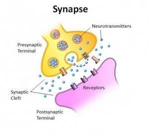
Chemical synapses are separated by a synaptic cleft which is filled with a matrix, which is there so that the post and pre-synaptic membranes adhere to each other.
The presynaptic element is the axon terminal, which contains synaptic vesicle –contain neurotransmitter- and secretory granules– contains soluble proteins. The membrane differentiations are dense accumulation of protein adjacent to and within the membrane. The proteins- shaped like pyramids- on the presynaptic side are the site of neurotransmitter release called active zone.
Postsynaptic density contains the neurotransmitter receptors which convert intercellular chemical signal into an intracellular signal–change in membrane potential.
Synaptic Arrangements
The Neuromuscular Junction
Most of what we know about the mechanisms of synaptic transmission was first established here and it has a clinical significance- disease, drugs and poisons.
Chemical synapses also occur between the axons of motor neurons on the spinal cord and skeletal muscle. The Neuromuscular Junction has one of the larges synapses in the body with a large number of active zones which are precisely aligned with a MOTOR PLATE– series of shallow folds,
Principles of Chemical Synaptic Transmission
Fast synaptic transmission at most CNS synapses is mediated by amino acids whereas Acetylcholine mediates fast synaptic transmission at ALL neuromuscular junctions.
Neurotransmitter synthesis and storage
GABA and the amines are made only by the neurons that release them. These neurons contain specific enzymes that synthesise the neurotransmitters from metabolic precursors.
The synthesizing enzymes for amino acids and amine neurotransmitters are transported to the axon terminal. Once synthesized in the cytosol of the axon terminal, the amino acid and amine neurotransmitters must be taken up by the synaptic vesicles.
TRANSPORTERS- special proteins embedded in the vesicle membrane that concentrate the neurotransmitter inside the vesicle
Secretory granules containing the peptide neurotransmitter bud of the Golgi apparatus and are carried to the axon terminal by axoplasmic transport.
Neurotransmitter Release
Depolarisation of the terminal membrane causes voltage gated calcium in the active zones to open.
There is a large inward driving force of CA2. At rest the calcium ion concentration is low and therefore calcium will flood the cytoplasm of the axon terminal as long as the calcium ions channels are open.
The vesicles release their contents through a process known as exocytosis and later recovered by the process of ENDOCYTOSIS and refilled with neurotransmitter.
Secretory granules release peptide neurotransmitters by exocytosis in a calcium-dependent fashion, but not as a typically the active zones. The release of peptide generally requires high-frequency trains of action potentials.
Neurotansmitter Receptors and Effectors
Transmitter Gated- Ion Channels (1)
Membrane spanning proteins consisting of 4/5 subunits that come together and form a pore, in the absence of a neurotransmitter the pore is closed. The neurotransmitter induces a conformational change.
EPSP A transient post-synaptic membrane depolarisation caused by the presynaptic release of neurotransmitter is called an excitatory post-synaptic potential
Synaptic activation of Ach-gated and glutamate- gated ion channels cause EPSP’s. Those permeable to Na+ and K+.
IPSP is a transient post-synaptic hyperpolarisation of the membrane caused by the presynaptic release of neurotransmitter is called inhibitory post-synaptic potential. Synaptic activations of glycine-gated or GABA gated ion channels cause an IPSP.
G-Protein Coupled Receptor (2)
Fast chemical synaptic transmission is mediated by amino acids and amine neurotransmitters acting on transmitter gated ion channels. However all three types of neurotransmitter can also have slower and longer lasting much more diverse post synaptic actions
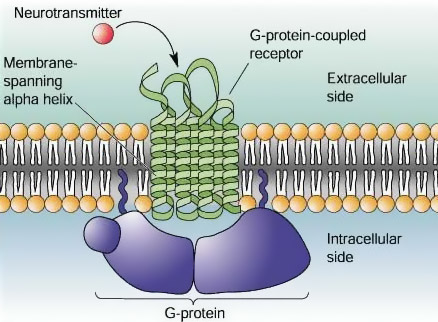
- Neurotransmitter molecules bind to receptor proteins embedded in the post synaptic membrane
- The receptor proteins activate small proteins called G-proteins that are free to move along the intracellular face of the post-synaptic membrane.
- The activated G-protein activate effector proteins
- G-protein coupled receptors are often referred to as metatrophic receptors- membrane receptors of eukaryotic cells that acts through a secondary messenger.
G-coupled receptors act as modulators, rather than directly elicit an EPSP or an IPSP
Autoreceptors (3)
Autoreceptors are a part of the post-synaptic density, and are presynaptic receptors that are sensitive to the neurotransmitter released by the presynaptic terminal are called autoreceptors.
Typically autoreceptors are G-protein coupled receptors that stimulate secondary messengers
A common effect is neurotransmitter release inhibition this allows the presynaptic membrane to regulate itself.
Neurotransmitter Recovery and Degradation
Reuptake occurs by the action of specific neurotransmitters transporter proteins located in the presynaptic membrane
Once inside the terminal the transmitters may be enzymatically destroyed or reloaded into synaptic vessels.
Enzymatic destruction occurs in the synaptic cleft. This is how acetylcholine is removed at the neuromuscular junction.


